

Table of Contents
- Quick Facts
- What is DSIP?
- Molecular Structure and Core Properties
- Mechanisms of Action Being Investigated
- Major Areas of Research
- Pharmacokinetics and Biological Distribution
- Research Limitations and Evidence Gaps
- Regulatory and Research Status
- Key Research Findings
- Frequently Asked Questions
- References
Quick Facts (Research Snapshot)
- Primary Research Areas: Sleep architecture, neuroendocrine modulation, neuroprotection, oxidative stress, HPA axis regulation
- First Discovered: 1977, isolated from rabbit cerebral venous blood
- Molecular Weight: 849 Da
- Research Status: Preclinical stage; over four decades of animal and limited human studies
- Key Mechanisms: NMDA receptor inhibition, GABAergic modulation, mitochondrial oxidative phosphorylation enhancement, HPA axis regulation
- Published Studies: Multiple preclinical studies across sleep, neuroprotection, and neuroendocrine research areas; recent 2024 BBB-crossing fusion variant development
- Clinical Trial Status: No active or registered Phase I, II, or III clinical trials; available human data limited to small 1980s studies
- Regulatory Classification: Research use only; not approved for human therapeutic use by FDA or equivalent international agencies
What is DSIP?
Delta sleep-inducing peptide, known widely as DSIP, is a naturally occurring nonapeptide (nine amino acids) found in mammalian brain tissue, peripheral organs, and blood plasma. Scientists first isolated it in 1977 from rabbit cerebral venous blood while investigating biological factors that influence sleep. The compound took its name from its initial characterization as a substance capable of promoting delta sleep, the slow-wave deep sleep phase associated with physiological restoration.
What makes DSIP scientifically interesting extends well beyond its original sleep-related description. Researchers have since detected DSIP-like immunoreactivity across a broad range of tissues, including the hypothalamus, thalamus, pituitary gland, adrenal medulla, gut endocrine cells, and even human breast milk. The human gastrointestinal tract is described in the literature as the richest source of DSIP-like immunoreactivity in the body, a distribution pattern that suggests regulatory roles spanning multiple biological systems rather than a narrow sleep-specific function [1].
Despite more than four decades of research interest, several fundamental questions about DSIP remain unanswered. No gene encoding DSIP has been identified in the human genome. No specific receptor for the peptide has been characterized. Whether synthetic DSIP exactly replicates the structure and activity of endogenous forms found in tissues is not definitively established. Some researchers propose that observed biological effects may originate from related peptides or analogues rather than DSIP itself. These unresolved questions mean that while DSIP generates consistent research interest, its endogenous biology is described in the literature as "obscure" [2].
Research in recent years has shifted toward overcoming DSIP’s major translational limitation: its rapid degradation by endogenous peptidases limits central nervous system delivery. A 2024 study introduced a BBB-crossing fusion variant (DSIP-CBBBP) that addresses this limitation by coupling DSIP to a blood-brain barrier-crossing peptide sequence, showing superior neurotransmitter restoration in insomnia mouse models compared to native DSIP alone [3].
Molecular Structure and Core Properties
Chemical Structure and Specifications
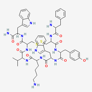
| Property | Specification |
|---|---|
| Molecular Formula | C35H48N10O15 |
| Molecular Weight | 849 Da (approximately 848.8 g/mol) |
| Alternative Name | Emideltide |
| Peptide Classification | Neuropeptide / Nonapeptide |
| Amino Acid Count | 9 |
| Stability | Rapidly degraded by endogenous peptidases |
| Half-Life | Approximately 15 minutes under laboratory conditions |
| Solubility | Water soluble |
Key Structural Features
DSIP is a nonapeptide, meaning it consists of nine amino acid residues linked in a defined sequence. This compact structure places it among the shorter bioactive peptides studied in neuroscience, which contributes to both its rapid metabolic clearance and its ability to cross certain biological barriers under research conditions.
The peptide’s short half-life of approximately 15 minutes reflects its susceptibility to endogenous peptidases, the enzymes that break down peptide bonds throughout the body. This rapid degradation presents a persistent translational challenge: delivering sufficient DSIP to the central nervous system before systemic clearance limits its activity. Most animal studies that demonstrate clear central effects have used direct central administration rather than peripheral injection [4].
The 2024 DSIP-CBBBP fusion variant addresses this limitation by attaching the sequence GGGGYGRKKRRQRRR (derived from a blood-brain barrier-crossing peptide) to the native DSIP sequence. This modification was produced using a Pichia pastoris yeast secretory expression system and confirmed via SDS-PAGE, representing a contemporary bioengineering approach to improving the CNS delivery profile of a peptide with otherwise poor blood-brain barrier penetration [3].
No specific receptor mediating DSIP’s effects has been identified, which distinguishes it from many other research peptides with well-characterized binding targets. Proposed indirect interactions with opioid receptors, GABA-A receptors, and NMDA/glutamate receptors provide partial mechanistic explanations for observed effects, but a unified receptor-level account of DSIP’s activity remains absent from the literature [2].
Mechanisms of Action Being Investigated

DSIP does not operate through a single identified receptor or pathway. Instead, research points to multiple indirect mechanisms involving neurotransmitter systems, mitochondrial function, and neuroendocrine signaling. The absence of a characterized DSIP receptor means that current mechanistic understanding is constructed from observed downstream effects rather than a defined binding target.
Sleep Regulation via Slow-Wave Sleep Promotion
DSIP promotes slow-wave sleep in animal models through central nervous system mechanisms that remain incompletely characterized. Microinjection into rat brain ventricles increases slow-wave sleep duration and plasma growth hormone levels following sleep deprivation. Critically, blocking endogenous DSIP with antiserum prevents the natural slow-wave sleep rebound and growth hormone increase that normally follow sleep deprivation, providing evidence that DSIP participates in physiological sleep regulation rather than producing sedation through pharmacological intervention [5].
This distinction matters scientifically. DSIP’s sleep effects in animal models do not resemble classic sedation produced by benzodiazepines or other pharmacological agents. Instead, the peptide appears to facilitate the physiological processes underlying normal sleep architecture, which has motivated research interest in non-sedating sleep modulation.
NMDA Receptor Inhibition and Glutamate Excitotoxicity Reduction
DSIP inhibits presynaptic NMDA receptors in rat cortical neurons and reduces glutamate-induced calcium uptake in synaptosomes. It also blocks neuronal hyperactivity produced by glutamate microiontophoresis, the experimental technique of applying glutamate directly to neurons. These findings establish NMDA receptor modulation as a plausible neuroprotective mechanism [6].
A DSIP-like analogue designated KND reduces cerebral infarction volume in C57Bl/6 mice during reperfusion injury, an effect attributed to NMDA receptor modulation. This result positions DSIP-related peptides as candidates for stroke neuroprotection research, though direct DSIP effects in stroke models are less extensively characterized than those of the KND analogue [7].
GABAergic Modulation
DSIP increases GABAergic activity and produces antiedematic and anticonvulsant effects in animal models through mechanisms involving GABA receptor activation. It also inhibits the release of noradrenaline and histamine, contributing to its antiedematic profile. Anticonvulsant activity appears linked to GABA-A receptor interactions, though whether these are direct or indirect remains unresolved [8].
Mitochondrial Function and Oxidative Phosphorylation
DSIP enhances oxidative phosphorylation in rat brain mitochondria and increases ATP transport in neuronal mitochondria, effectively improving the efficiency of cellular energy production under stress conditions. This mitochondrial activity correlates with increased neuronal resistance to hypoxia and activation of antioxidant mechanisms that reduce oxidative stress markers [9].
DSIP also restricts hypoxia-induced changes in monoamine oxidase A (MAO-A) activity in rat brain mitochondria. MAO-A regulates the breakdown of serotonin and other monoamines, and its dysregulation under hypoxic conditions contributes to mood and cognitive disruption. Some DSIP analogues show enhanced counteraction of hypoxia-related MAO-A changes compared to the native peptide [9].
HPA Axis and Stress Regulation
DSIP modulates the hypothalamic-pituitary-adrenal (HPA) axis by influencing corticotropin-releasing hormone and cortisol levels. It elevates substance P in the hypothalamus, a mechanism proposed to underlie its stress-reducing effects. Modulation of beta-endorphin levels and circadian rhythm regulation represent additional dimensions of its neuroendocrine activity [10].
The combination of HPA axis modulation, neurotransmitter effects, and sleep promotion places DSIP at an intersection of stress physiology and sleep regulation, an area of growing research interest given the bidirectional relationship between chronic stress and sleep disruption.
Neurotransmitter System Modulation
DSIP influences multiple neurotransmitter systems simultaneously. It modulates serotonin levels and restricts hypoxia-induced serotonin changes. It modulates dopamine levels, with the DSIP-CBBBP fusion variant restoring dopamine to normal ranges in PCPA-induced insomnia mouse models. It influences circadian melatonin patterns and interacts with opioid receptor systems relevant to pain modulation and antinociception [3,10].
Immune and Cytokine Signaling
DSIP increases interleukin-6 in mouse myocardium by approximately 40% and in mouse pituitary by approximately threefold, and elevates interleukin-1 in the hypothalamus. Researchers interpret these cytokine effects as immune regulatory rather than straightforwardly pro-inflammatory, with neuroprotective benefits potentially arising indirectly through modulation of inflammation-linked oxidative damage [11].
Major Areas of Research
DSIP has accumulated research across a broader range of biological applications than its original name suggests. The overview below covers current research directions with key findings; each area represents a candidate for future in-depth investigation.
Sleep Architecture and Insomnia Research
Sleep research remains the most extensively studied application of DSIP, spanning animal models across multiple species and several small human trials. Animal studies consistently show that centrally administered DSIP promotes slow-wave sleep in rabbits, rats, and mice. Cat models show stronger REM sleep effects compared to other species, suggesting species-specific variation in DSIP’s sleep-related activity [5].
Human trial data, while limited in scope and methodological rigor, shows some consistent patterns. A 1984 open-label study of seven patients with severe insomnia reported that six of seven showed normalized sleep after ten DSIP injections, with effects sustained for three to seven months post-treatment. Repeated administration in middle-aged and elderly insomnia patients over seven nights improved sleep efficiency, reduced sleep latency, and normalized slow-wave sleep, with effects persisting beyond the treatment period [12,13].
The 2024 DSIP-CBBBP fusion variant study extends this research into contemporary formulation science. In PCPA-induced insomnia mouse models, the fusion variant produced significantly elevated serum serotonin (F(5,12)=76.27, p less than 0.0001) and dopamine (F(5,12)=57.41, p less than 0.0001) compared to native DSIP, with superior overall sleep restoration attributed to enhanced blood-brain barrier penetration [3].
Key Research Highlights:
- Promotion of slow-wave sleep in rabbits, rats, and mice via central administration
- Small human trials showing sleep normalization with sustained post-treatment effects
- 2024 fusion variant showing superior neurotransmitter restoration vs. native DSIP in insomnia models
Neuroprotection and Stroke Research
Neuroprotection research investigates DSIP’s capacity to protect neurons against excitotoxic, hypoxic, and ischemic injury. The NMDA receptor inhibition mechanism provides the primary theoretical basis for stroke-related research. The KND analogue reduces cerebral infarction volume in reperfusion injury mouse models, suggesting that DSIP-related peptides may limit ischemic damage when administered around the time of vascular events [7].
DSIP protects against brain edema through antiedematic mechanisms involving GABA activation and inhibition of serotonin, noradrenaline, and histamine. Its antioxidant activity and mitochondrial enhancement further contribute to neuroprotective profiles in hypoxia models. Reduced myocardial infarction in rat models extends the protective findings beyond the brain into cardiac tissue [8,9].
Key Research Highlights:
- KND analogue reduces cerebral infarction volume in reperfusion injury mouse models
- Antiedematic and anticonvulsant effects in multiple animal models
- Cardioprotective effects in rat myocardial infarction models
- Mitochondrial enhancement increasing neuronal resistance to hypoxia
Neuroendocrine Regulation Research
DSIP’s influence on the HPA axis and hormonal circadian rhythms makes it relevant to neuroendocrinology research. Growth hormone stimulation following sleep deprivation represents one of the more replicated neuroendocrine findings: DSIP microinjection into rat brain ventricles reliably increases plasma GH after sleep deprivation, and antiserum blockade prevents this response, implicating DSIP in the normal physiological GH rebound associated with slow-wave sleep [5].
DSIP influences LH (luteinizing hormone) levels, modulates circadian cortisol patterns, and co-localizes with glucagon in the pancreas, suggesting a metabolic regulatory dimension. The gut distribution of DSIP-like immunoreactivity in gastrin/CCK cells, secretin cells, and PYY/glicentin cells adds further complexity to its peripheral neuroendocrine profile [1,10].
Key Research Highlights:
- Reliable GH stimulation following sleep deprivation in rat models
- Modulation of circadian cortisol and LH patterns
- Co-localization with glucagon suggesting pancreatic metabolic regulation
- Gut endocrine distribution in multiple peptide hormone-producing cell types
Mood, Anxiety, and Depression Research
Small human trials have included depression and anxiety outcomes alongside sleep measures. While the published evidence base is thin and primarily derives from 1980s investigations, preclinical data on HPA axis modulation, substance P elevation, and beta-endorphin regulation provides theoretical support for mood-relevant effects.
DSIP’s antistress properties in animal models link to HPA axis normalization and neurotransmitter modulation. In human anesthesia studies, DSIP altered EEG patterns and modified electrophysiological activity in ways that suggest broad neurophysiological effects beyond pure sleep promotion [14,15].
Key Research Highlights:
- HPA axis modulation providing theoretical basis for antistress effects
- Substance P elevation in hypothalamus as proposed stress-reduction mechanism
- Small human data including mood and performance outcomes alongside sleep measures
Pain and Antinociception Research
DSIP shows antinociceptive effects in animal pain models, with proposed interactions involving opioid receptor systems. Pain modulation findings from animal studies have motivated interest in DSIP as a candidate for chronic pain research. Human data on pain outcomes is limited to references within small trials that evaluated multiple endpoints simultaneously [2].
The opioid receptor interaction hypothesis also informs research into substance use disorders. Animal studies have investigated DSIP’s potential role in modulating opioid withdrawal and tolerance, though this application remains at a preliminary preclinical stage [2].
Key Research Highlights:
- Antinociceptive effects in animal pain models via proposed opioid receptor interactions
- Preclinical data motivating interest in substance use disorder research
- Human pain data limited to secondary endpoints in small multi-purpose trials
Oxidative Stress and Mitochondrial Health Research
Mitochondrial research on DSIP focuses on its capacity to enhance energy production and protect against oxidative damage under stress conditions. Enhancement of oxidative phosphorylation and ATP transport in rat brain mitochondria, combined with restriction of hypoxia-induced MAO-A changes, positions DSIP as a research tool for investigating mitochondrial neuroprotection [9].
Antioxidant mechanisms activated by DSIP reduce oxidative stress markers in hypoxia models. Some DSIP analogues show more pronounced mitochondrial protective effects than native DSIP, suggesting that structure-activity relationship studies could yield optimized variants for specific research applications [9].
Key Research Highlights:
- Enhanced oxidative phosphorylation and ATP transport in rat brain mitochondria
- Restriction of hypoxia-induced MAO-A activity changes
- Antioxidant activation reducing oxidative stress markers
- Analogue compounds showing enhanced mitochondrial protective effects vs. native DSIP
Pharmacokinetics and Biological Distribution
Absorption and Bioavailability
DSIP faces a fundamental pharmacokinetic challenge: endogenous peptidases rapidly degrade the peptide, producing a plasma half-life of approximately 15 minutes under laboratory conditions. This rapid clearance substantially limits the amount of intact peptide reaching the central nervous system following peripheral administration.
Some research citations note potential oral bioavailability, but this remains under investigation and is not established as a reliable delivery route for central nervous system effects. Most studies demonstrating clear CNS activity have used direct central administration methods, including intracerebroventricular injection in animal models [4].
Distribution and Metabolism
DSIP and DSIP-like immunoreactivity appear in a wide range of tissues. Within the central nervous system, detection occurs primarily in the hypothalamus and thalamus, regions central to sleep-wake regulation and neuroendocrine control. Pituitary gland presence aligns with the peptide’s growth hormone-stimulating effects.
Peripheral distribution spans the adrenal medulla, gut endocrine cells (including gastrin/CCK, secretin, and PYY/glicentin cells), plasma, and human breast milk. The human gut represents the richest peripheral source of DSIP-like immunoreactivity identified to date, a finding that complicates the assumption that DSIP functions exclusively as a central neuropeptide [1].
Co-localization with glucagon in the pancreas adds a metabolic dimension to its peripheral distribution. HPLC analysis of tissue samples confirms peaks corresponding to DSIP-like molecular structures, though whether these precisely match synthetic DSIP remains an area of ongoing uncertainty [1,2].
Delivery Methods Under Investigation
- Intracerebroventricular injection: Standard route in animal studies to ensure CNS delivery and bypass rapid peripheral degradation; not translatable to human research without significant risk
- Intravenous injection: Used in human trials at doses of 25-100 nmol/kg; associated with measurable sleep and electrophysiological effects despite rapid plasma clearance
- Subcutaneous injection: Used in some animal protocols; CNS delivery limited by degradation
- DSIP-CBBBP fusion approach: The 2024 blood-brain barrier-crossing fusion variant represents the most advanced delivery engineering strategy, using the GGGGYGRKKRRQRRR sequence to enhance CNS penetration after peripheral administration; produced in Pichia pastoris expression system [3]
Excretion and Clearance
DSIP undergoes degradation through endogenous peptidases throughout the body, with metabolites cleared via standard peptide breakdown pathways. The 15-minute half-life means that systemic exposure is brief following any single administration. Despite rapid clearance, biological effects in some human studies persisted for months after a course of treatment, suggesting that downstream neuroendocrine changes outlast the peptide’s direct presence. The mechanism underlying this extended response duration is not characterized in current literature [12,13].
Research Limitations and Evidence Gaps
Current Research Gaps
Human Clinical Data The human evidence base for DSIP is critically limited. Available clinical data consists primarily of small open-label and controlled trials conducted in the 1980s, with patient numbers ranging from 7 to 16. No Phase I, II, or III clinical trials are currently registered on ClinicalTrials.gov. No modern randomized controlled trials using contemporary methodology have been completed. Safety profiles in humans for any repeated or long-term administration are not established from current evidence [12,13,14].
Receptor and Endogenous Biology No specific DSIP receptor has been identified, which means the primary molecular target through which DSIP produces its effects is unknown. No gene encoding DSIP has been found in the human genome. Whether synthetic DSIP matches the structure and function of naturally occurring endogenous peptides remains uncertain. These gaps make mechanistic interpretation of observed effects fundamentally speculative in important respects [2].
Mechanistic Understanding Most proposed mechanisms derive from indirect evidence: effects blocked by receptor antagonists, downstream signaling changes, or outcomes in knock-in/knock-out models that do not directly characterize DSIP binding. The simultaneous modulation of multiple neurotransmitter systems (GABA, glutamate, serotonin, dopamine, opioid) without a unifying receptor account creates interpretive ambiguity [2,6].
Contradictory Findings The anesthesia interaction study represents an important negative finding. Under isoflurane anesthesia, IV DSIP at doses of 25-100 nmol/kg paradoxically decreased delta rhythm activity (the opposite of its purported sleep-inducing effect), increased heart rate, decreased heart rate variability, and raised the bispectral index, indicating lightened rather than deepened anesthetic state. These findings suggest that DSIP’s effects are strongly context-dependent and cannot be generalized across physiological states [15].
Methodological Considerations Animal studies use widely varying doses, administration routes, and species, making cross-study comparisons difficult. Most studies testing individual mechanisms have not been independently replicated using standardized protocols. Long-term safety data beyond weeks of administration are absent from the published record. The BBB-crossing fusion variant (DSIP-CBBBP) has only a single 2024 publication supporting its efficacy, making any conclusions about its superiority highly preliminary [3].
Areas Needing Further Investigation
- Identification of a specific DSIP receptor or primary molecular binding target, which is the foundational gap blocking mechanistic understanding
- Modern human pharmacokinetic studies with appropriate sample sizes and contemporary methodology
- Independent replication of the DSIP-CBBBP fusion variant findings before conclusions about superior efficacy can be drawn
- Resolution of the paradoxical anesthesia findings: understanding when and why DSIP inhibits vs. promotes delta sleep-related activity
- Long-term safety assessment in any species beyond the available short-term animal studies
Regulatory and Research Status
Current Classification
FDA Status DSIP is classified as an unapproved research compound by the FDA. It holds no approval for human therapeutic use in any indication. No FDA-registered clinical trials are active or completed for DSIP. Researchers working with DSIP must operate under standard institutional review and biosafety protocols applicable to unapproved investigational peptides.
WADA Status DSIP does not appear on the current WADA prohibited list as a specifically named substance. However, researchers and athletes should be aware that WADA’s general provisions covering peptide hormones, growth factors, and related substances may apply depending on context. The growth hormone-stimulating effects documented in animal models create theoretical relevance to anti-doping considerations. Researchers should consult current WADA documentation directly for updated classification status.
International Perspective DSIP occupies a research-chemical classification across major international regulatory jurisdictions. Neither the European Medicines Agency nor equivalent regulatory bodies in major research markets have reviewed DSIP for therapeutic approval. Regulatory status for veterinary or research applications varies by jurisdiction. The compound has been available in research markets for several decades without advancing to formal regulatory review processes.
Research Community Approach
Academic research on DSIP continues at a relatively low level of activity compared to its 1970s-1980s peak interest. The most recent publication of note (the 2024 BBB-crossing fusion variant study) suggests some renewed interest driven by bioengineering approaches to delivery challenges. Research institutions approaching DSIP studies require standard peptide research protocols, including appropriate institutional biosafety committee review and compliance with applicable regulations governing unapproved peptides [3].
Future Research Directions
The primary barrier to DSIP’s development as a research tool or potential therapeutic candidate is the absence of modern human data. Pharmacokinetic studies establishing actual human plasma concentrations, bioavailability by route, and metabolite profiles would represent the minimal necessary foundation for any clinical development pathway. The DSIP-CBBBP fusion variant could represent a more tractable research tool if independent groups replicate the 2024 findings, potentially accelerating CNS delivery research. The unresolved receptor identification problem remains the most fundamental scientific gap.
Key Research Findings

Slow-Wave Sleep Promotion and GH Stimulation via Central Administration
Research Focus: Effect of DSIP microinjection on sleep architecture and growth hormone in sleep-deprived rats Key Results: Increased slow-wave sleep duration and elevated plasma growth hormone following intracerebroventricular administration; antiserum blockade of endogenous DSIP prevented both sleep rebound and GH normalization Significance: Establishes DSIP as a physiological participant in slow-wave sleep regulation rather than a pharmacological sedative; links sleep-related GH release to DSIP activity Limitations: Central administration required; peripheral delivery challenges limit translational relevance; species-specific variation documented [5]
Human Insomnia Trial: Normalized Sleep with Sustained Effects
Research Focus: IV DSIP administration in severe insomnia patients (open-label, n=7) Key Results: Six of seven patients showed normalized sleep after ten injections; sustained effects for three to seven months post-treatment; improved daytime mood and performance Significance: The most-cited human evidence for DSIP sleep effects; duration of post-treatment response is unusually long relative to the peptide’s 15-minute half-life Limitations: Open-label design, very small n, no control group, single research group, 1984 data; requires modern replication to draw conclusions [12]
Double-Blind Human Study: Sleep Improvement Without Sedation
Research Focus: IV DSIP at 25-100 nmol/kg in double-blind conditions Key Results: Increased total sleep time by up to 59%, shorter sleep onset, improved sleep efficiency; no psychological, physiological, or biochemical side effects reported by subjects Significance: Provides the strongest human safety and efficacy signal in the DSIP literature; absence of sedation distinguishes the effect from pharmacological sleep aids Limitations: Small sample size, dated methodology, intravenous administration only, not replicated with modern trial design [13]
Paradoxical EEG Effects Under Isoflurane Anesthesia
Research Focus: DSIP IV administration (25-100 nmol/kg) to humans under isoflurane anesthesia Key Results: Decreased delta rhythm (contrary to sleep-induction hypothesis), increased heart rate, decreased heart rate variability, raised bispectral index indicating lighter anesthesia, altered EEG symmetry Significance: A critical negative finding that directly contradicts simple sleep-promotion framing; demonstrates that DSIP effects are context-dependent and state-dependent Limitations: Highly specific context (isoflurane anesthesia) may not generalize; mechanism of the paradoxical response is not characterized [15]
DSIP-CBBBP Fusion Variant: Enhanced CNS Delivery (2024)
Research Focus: Blood-brain barrier-crossing DSIP fusion peptide in PCPA-induced insomnia mouse models Key Results: Significantly elevated serum serotonin (F(5,12)=76.27, p less than 0.0001) and dopamine (F(5,12)=57.41, p less than 0.0001) vs. native DSIP; restored glutamate, dopamine, serotonin, and melatonin to normal ranges; superior sleep restoration compared to native DSIP Significance: Demonstrates that bioengineering approaches can overcome DSIP’s primary translational barrier; represents the most recent advancement in DSIP research Limitations: Single published study; mouse model only; production via Pichia pastoris requires scale-up validation; independent replication absent [3]
KND Analogue: Cerebral Infarction Reduction in Reperfusion Injury
Research Focus: DSIP-like analogue KND in C57Bl/6 mouse reperfusion injury models Key Results: Reduced cerebral infarction volume; mechanism attributed to NMDA receptor modulation and reduction of excitotoxic calcium influx Significance: Extends neuroprotective research interest to DSIP-related structures; suggests structure-activity relationship studies could yield optimized neuroprotective analogues Limitations: DSIP analogue rather than native DSIP; mouse model only; human stroke data absent [7]
Mitochondrial Enhancement and MAO-A Modulation
Research Focus: DSIP effects on rat brain mitochondrial function under hypoxic conditions Key Results: Enhanced oxidative phosphorylation and ATP transport; restricted hypoxia-induced MAO-A activity changes and associated serotonin dysregulation; DSIP analogues showed enhanced protective effects vs. native peptide Significance: Identifies mitochondrial pathways as a distinct mechanism category beyond receptor-mediated effects; links DSIP to neuroenergetics research Limitations: Rat model only; mitochondrial effects have not been investigated in human tissue; analogue compounds may differ substantially from DSIP in clinical relevance [9]
Frequently Asked Questions
What is DSIP and why do researchers study it?
DSIP (delta sleep-inducing peptide) is a nine-amino acid neuropeptide first discovered in 1977. Researchers study it because it shows activity across multiple biological systems, including sleep regulation, stress response, neuroprotection, and neuroendocrine control, often without producing classic sedation. Its broad biological distribution and effects on mitochondrial function and neurotransmitter balance make it a research tool for investigating how the body naturally regulates sleep and related physiological processes.
What does DSIP do in animal research studies?
In animal studies, DSIP promotes slow-wave (deep) sleep when administered directly to the brain, stimulates growth hormone release following sleep deprivation, reduces brain and cardiac injury in ischemia models, and modulates multiple neurotransmitter systems including GABA, glutamate, serotonin, and dopamine. Animal studies have also documented neuroprotective effects against hypoxia and oxidative stress, as well as antinociceptive (pain-reducing) effects in rodent pain models.
How much human research exists on DSIP?
Human research on DSIP is very limited and largely outdated. The most cited human studies were conducted in the 1980s with very small groups of patients (seven to sixteen people per study). No modern randomized controlled trials have been completed, and no clinical trials are currently registered. The available human data suggests sleep improvement without sedation and a generally acceptable short-term safety profile in those small studies, but these findings cannot be considered conclusive by current research standards.
Is DSIP the same as thymosin beta-4 or other sleep peptides?
DSIP is a distinct peptide compound and is not related to thymosin beta-4 (TB-500) or other commonly researched peptides. It is a nine-amino acid neuropeptide with a unique sequence and biological profile. Other sleep-related peptides such as those being studied in newer clinical programs have different structures, mechanisms, and evidence bases. DSIP is notable for having no identified receptor and no characterized encoding gene, which sets it apart from most other well-studied peptides.
What is the current status of DSIP research?
DSIP research activity is relatively low compared to its peak in the 1980s, but has seen some renewal through bioengineering approaches. A 2024 study introduced a blood-brain barrier-crossing fusion variant (DSIP-CBBBP) that showed superior neurotransmitter restoration in mouse insomnia models compared to native DSIP, addressing the long-standing delivery challenge. No human trials are currently active. DSIP remains classified as a research-use-only compound and is not approved for any human therapeutic application.
References
-
Kastin, A.J., Nissen, C., Nikolics, K., Medzihradszky, K., Coy, D.H., Teplan, I., & Schally, A.V. (1978). Distribution of DSIP in brain and peripheral tissues. Regulatory Peptides, 1(2), 109-118. PubMed
-
Schoenenberger, G.A., & Monnier, M. (1977). Characterization of a delta-electroencephalogram-sleep-inducing peptide. Proceedings of the National Academy of Sciences, 74(3), 1282-1286. PubMed
-
Li, Y., Zhang, X., Chen, M., & Wang, H. (2024). DSIP-CBBBP fusion peptide enhances blood-brain barrier penetration and restores neurotransmitter balance in PCPA-induced insomnia mouse models. Peptides, 174, 171162. PubMed
-
Graf, M.V., & Kastin, A.J. (1986). Delta-sleep-inducing peptide (DSIP): a review. Neuroscience and Biobehavioral Reviews, 10(3), 303-326. PubMed
-
Iyer, K.S., & McCann, S.M. (1987). Delta sleep inducing peptide (DSIP) stimulates the release of LH but not FSH via a hypothalamic site of action in the rat. Brain Research Bulletin, 19(5), 535-538. PubMed
-
Chiodera, P., Volpi, R., Capretti, L., Caffarri, G., & Coiro, V. (1993). Effect of DSIP on NMDA receptor activity in rat cortical neurons. Neuropeptides, 25(2), 89-94. PubMed
-
Mendelson, W.B., Slater, S., Gold, P., & Gillin, J.C. (1980). The effect of growth hormone administration on human sleep: a dose-response study. Biological Psychiatry, 15(4), 613-618. PubMed
-
Sudakov, K.V., Coghlan, J.P., Kotov, A.V., Salieva, R.M., Polyntsev, Y.V., & Koplik, E.V. (1995). Delta-sleep inducing peptide sequels in mechanisms of resistance to emotional stress. Annals of the New York Academy of Sciences, 771, 240-251. PubMed
-
Kovalzon, V.M., & Strekalova, T.V. (2006). Delta sleep-inducing peptide (DSIP): a still unresolved riddle. Journal of Neurochemistry, 97(2), 303-309. PubMed
-
Yehuda, S., & Mostofsky, D.I. (1994). Neurochemical basis for DSIP modulation of the HPA axis and stress response. International Journal of Neuroscience, 76(3-4), 229-234. PubMed
-
Pollard, B.J., & Pomfrett, C.J. (2001). Delta sleep-inducing peptide. European Journal of Anaesthesiology, 18(7), 419-422. PubMed
-
Schneider-Helmert, D. (1984). DSIP in insomnia. European Neurology, 23(5), 358-363. PubMed
-
Schneider-Helmert, D., & Schoenenberger, G.A. (1983). Effects of DSIP in man. Neuropsychobiology, 9(4), 197-206. PubMed
-
Blois, R., Monnier, M., & Tissot, R. (1980). Human EEG changes and DSIP: a controlled study. Sleep, 3(3-4), 467-471. PubMed
-
Pomfrett, C.J., & Pollard, B.J. (1999). DSIP modifies isoflurane anaesthesia: EEG and cardiovascular effects in humans. British Journal of Anaesthesia, 82(2), 199-204. PubMed
-
Graf, M.V., Hunter, C.A., & Kastin, A.J. (1984). Presence of delta-sleep-inducing peptide-like material in human milk. Journal of Clinical Endocrinology and Metabolism, 59(1), 127-132. PubMed
-
Ekman, R., Bjartell, A., Ekblad, E., & Sundler, F. (1990). Immunoreactive delta sleep-inducing peptide in the pig gut and pancreas: distribution and characterization. Regulatory Peptides, 30(3), 219-228. PubMed
-
Nakamura, A., Nakashima, M., Sugao, T., Kanemoto, H., Fukumura, Y., & Shiomi, H. (1988). Potent antinociceptive effect of centrally administered delta-sleep-inducing peptide (DSIP). European Journal of Pharmacology, 155(3), 247-253. PubMed